Medical Retina
Diabetic Retinopathy Treatment & Prevention
Welcome to Bansal Eye Hospital. BEH is the pioneer eye hospital in North India and one of the Best Eye Hospital in Chandigarh. Bansal Eye Hospital is empanelled with Haryana Government, Himachal Government, Punjab Government, ESIC- Himachal Pradesh, CGHS, ECHS for Lasik Laser Eye Surgery, Specs Removal Surgery, C3R Surgery, Cataract (Safed Motia or Motiabind) Surgery, Diabetic Retinopathy, Glaucoma (kala motia) and other eye related treatment and surgery.
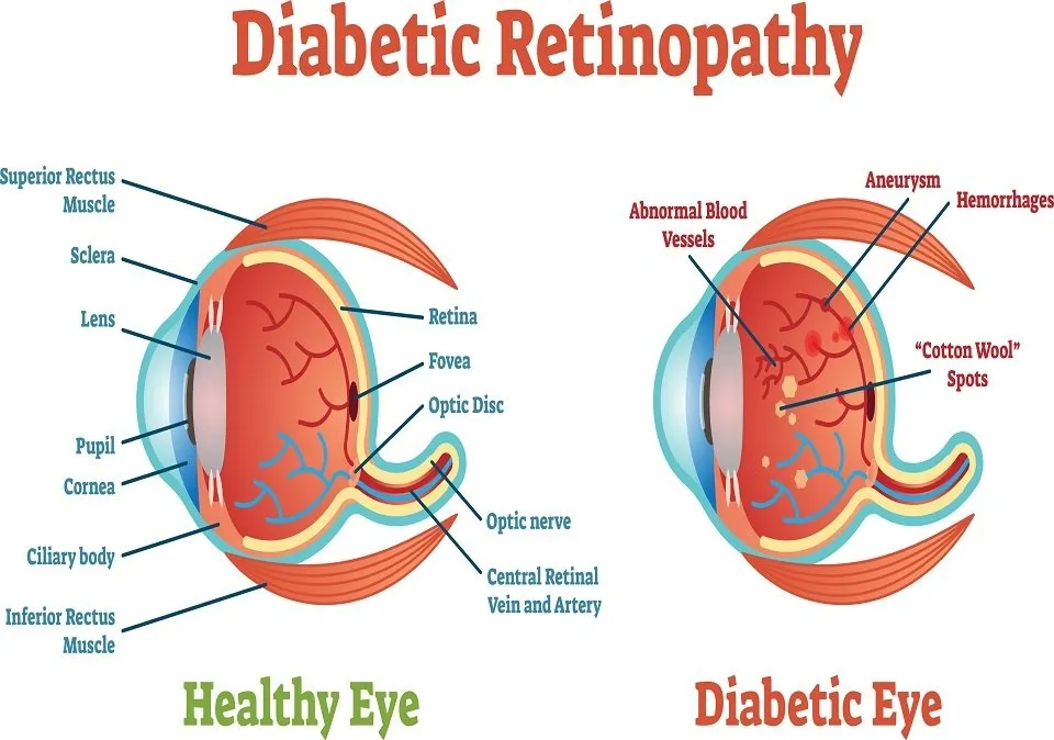
Retina is the photosensitive layer of the nerve cells lining the inside of the eye. It is affected in some hereditary disorders, diabetes, hypertension, blood disorders, injuries, vascular occlusions, aging and lot of other conditions. Cases with retinal disorders feel painless medical-retinarapid loss of vision.

Bansal Eye Hospital is fully equipped to diagnose and manage all kind of retinal disorders. We are equipped to manage patients with diabetic retinopathy, diabetic maculopathy, branch retinal vein occlusion (BRVO) and central retinal vein occlusion (CRVO), choroidal neovascular membranes (CNVM), eales disease and retinal vasculitis, retinal breaks and lattice degeneration with holes, Central serous chorioretinopathy (CSCR,) proliferative vitreo-retinopathies and many other retinal disorders.
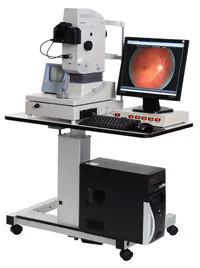
Digital fundus fluorescein angiography (STEREO), visual fields and laser are available for management of these cases. A Diode laser is available for anterior and posterior segment with endo photo coagulation attachment.
What Is Diabetic Retinopathy?
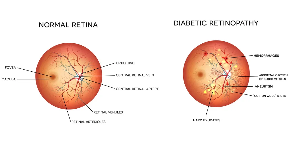
Diabetic Retinopathy, a complication of diabetes, is caused by changes in the blood vessels of the retina, the light sensing nerve layer at the back of the eye. These damaged blood vessels leak fluid or blood, and develop fragile brush like branches and scar tissue. The images which the retina sends to the brain BEHome blurred, distorted or partially blocked.
Who gets Diabetic Retinopathy?


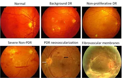
Diabetic retinopathy Stages
Mild Nonproliferative Retinopathy. At this stage, microaneurysms occur.
Moderate Nonproliferative Retinopathy. This stage is when blood vessels that nourish the retina are blocked.
Severe Nonproliferative Retinopathy.
Proliferative Retinopathy.
Diabetic Retinopathy
Types, Causes, Symptoms, Stage & Diagnosis
Type of Diabetic Retinopathy
Background retinopathy :-
Background retinopathy is an early state of diabetic retinopathy. In this stage, fine blood vessels within the retina BEHome narrowed or obstructed while others enlarge to form balloon-like sacs. These altered vessels leak blood and fluid, causing the retina to swell or form deposits called exudates.
Sight is usually not seriously affected. It can, however lead to more advanced sight-threatening stages, and for this reason is considered a warning sign. In some cases, the leaking fluid collects in the macula, the central part of the retina which is responsible for detailed vision, such as reading. This problem is called macular oedema or diabetic maculopathy. Reading and close work may BEHome more difficult BEHause of this condition.
Proliferative retinopathy :-
Proliferative retinopathy is an advanced stage. It describes the changes that occur when new, abnormal blood vessels begin to grow on the surface of the retina or the optic nerve. These new blood vessels, called neovascularisation, have weaker walls and may rupture and bleed into the vitreous, the clear gel like substance that fills the centre of the eye.
This leaking blood can cloud the vitreous and partially block the light through the pupil towards the retina, causing blurred and distorted images. These abnormal blood vessels frequently grow scar like tissue with them which may pull the retina away from its normal position at the back of the eye (detached retina.
Causes and Symptoms

The cause of diabetic retinopathy is not completely understood. However, it is known that diabetes damages small blood vessels in various parts of the body. The damage to the tiny blood vessels in the retina occurs when the blood sugar levels come down rapidaly.
Every fluctuation is thus dangerous.Diabetic retinopathy is a painless condition. It is not associated with redness or watering or irritation of the eye. One can have background diabetic retinopathy for a long time without any effect on vision. Thus changes in the eye can go unnoticed unless detected by a medical eye examination. Gradual painless blurring of vision may occur if macular oedema is present. A sudden loss of vision can occur if bleeding occurs inside the eye with proliferative retinopathy. This severe form of diabetic retinopathy requires immediate medical attention.

Detection & Diagnosis
Comprehensive medical eye examination
Fundus
photography
Fundus fluorescein
angiography
Laser Surgery

The small laser scars reduce abnormal blood vessel growth (neovascularisation) and help bond the retina to the back of the eye.Laser treatment is planned to take care of the existing lesions in retina. One or more than one session, spread over a week or so may be necessary. Laser are not a substitute for adequate control of diabetes. It may take three to six months for full effect of the laser therapy to be evident. Laser treatment can be repeated if required.If diabetic retinopathy is detected early, photocoagulation by laser surgery retards vision loss.
Even in the more advanced stages of the disease (proliferative retinopathy), it reduces the chance of severe visual impairment. The laser treatment prevents further loss of vision rather than restore the vision. It is therefore important to note, the the ideal time to start the treatment is when the vision is still normal.
Cost of Diabetic Retinopathy in Haryana?
Now the exact cost of C3R Eye LASIK and other Laser eye procedures that we have at our LASIK centre, Please fill the form or Call Our Customer Care.
Vision Loss is Largely Preventable
Early detection of diabetic retinopathy is the best protection against loss of vision. It is important to remember that diabetic retinopathy may be present without any symptoms. People with diabetes should get fundus (retina) examined by an ophthalmologist at least once a year. More frequent medical eye examinations may be necessary, once diabetic retinopathy has been diagnosed. In most cases, with careful monitoring, the ophthalmologist can begin treatment before sight is affected.
